|
|
  Laboratory of Mathematical Methods of Image Processing Chair of Mathematical Physics |
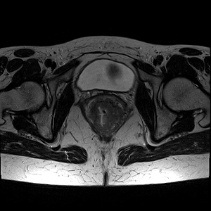
Biomedical Imaging — MRITumor segmentationWithin the work that was funded by RFBR, CNPq and MOST according to the research project 19-57-80014 (BRICS2019-394) we collected a dataset of annotated pelvic T2 3D MRI images with tumors. On each 3D image, a surgeon selected a slice with the maximum area of the tumor on the axial section, after which a polygonal mask corresponding to the tumor was created. All images in the dataset are anonymized.
|
|||||||